Mohs Micrographic Surgery
Mohs surgery in Plano, Texas
Skin cancer is what we do. Our three board-certified and fellowship-trained surgeons remove it with Mohs micrographic surgery, reading 100% of your margins in our own laboratory while you wait, with cure rates up to 99%.
What is Mohs surgery?
Mohs micrographic surgery is the most precise way to remove a skin cancer. Pioneered by Dr. Frederick Mohs and refined over nearly a century, it is today's gold standard for treating most skin cancers: the tumor is removed one thin layer at a time, and every margin is read under the microscope before you go home.
Physician-owned and operated · Physician referrals welcome · Most insurance accepted · Serving Dallas and all of North Texas · 6100 Windhaven Parkway, Plano, TX 75093




What sets Mohs apart
The gold standard for skin cancer treatment with up to 99% cure rate.
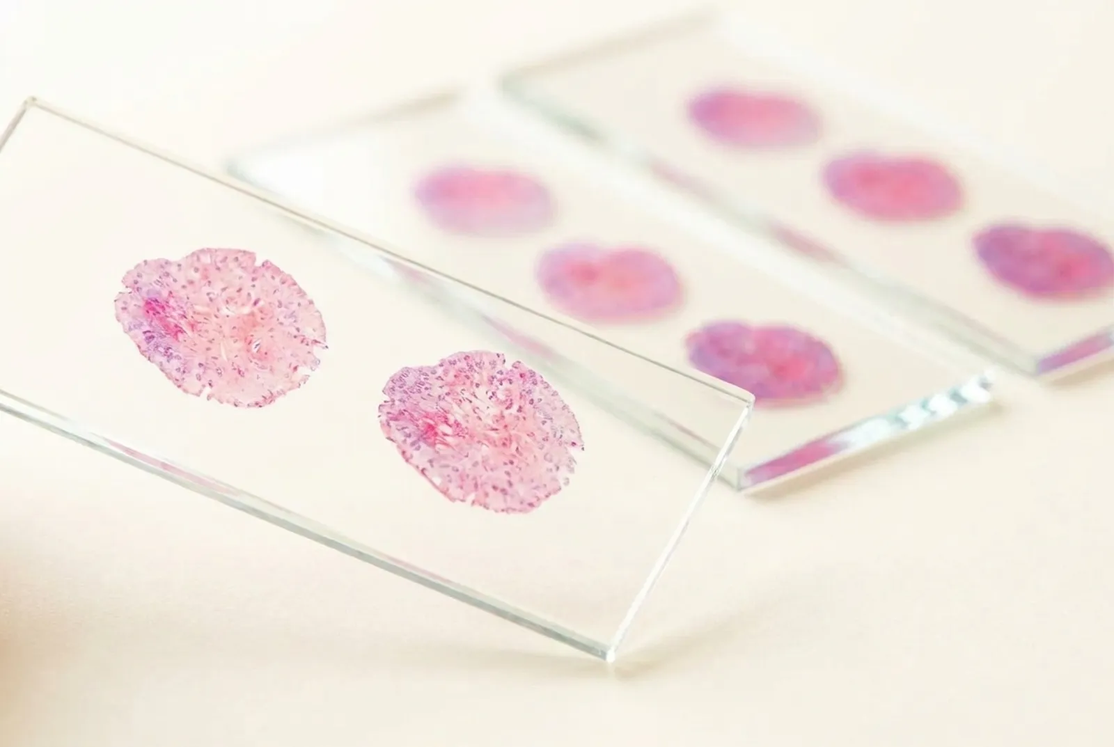
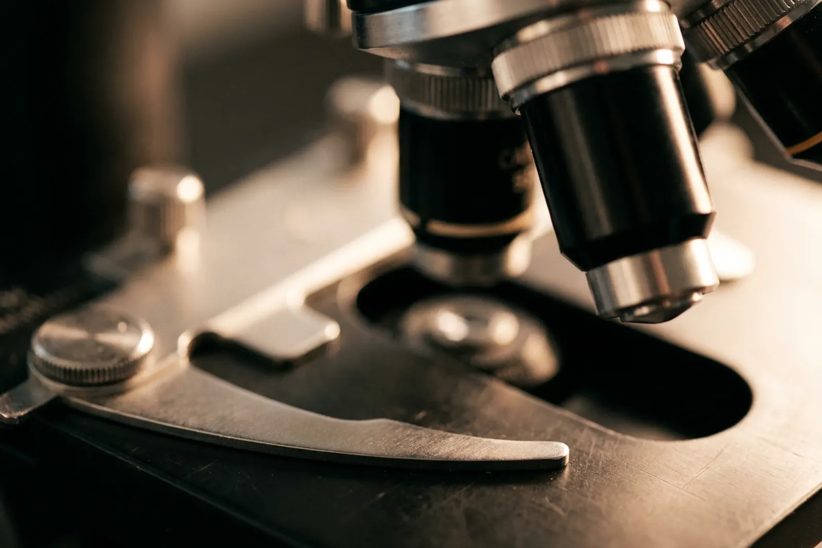
What sets Mohs apart is simple. Instead of removing a margin of healthy skin and waiting days for a lab to report whether the edges were clear, your surgeon examines 100% of the margin under the microscope while you wait. If any cancer remains, its exact location is mapped and only that spot is removed — the cancer is traced to its roots, and nothing healthy is taken unnecessarily.
Mohs surgery pairs the surgeon with dedicated laboratory staff and an on-site laboratory, where your tissue is frozen, sectioned, stained, and read within the hour it is removed. This practice is built around exactly that. The stages of a Mohs day take time because the microscope — not estimation — decides when your surgery is finished.
The result is the treatment's defining combination: cure rates up to 99% for previously untreated cancers, the most healthy skin preserved, and an answer before you leave that the cancer is gone.
- Up to 99% cure rate for previously untreated cancers
- Tissue-sparing technique preserves maximum healthy skin
- Complete microscopic examination during surgery
- Same-day results - know cancer is gone before you leave
- Optimal cosmetic outcomes with minimal scarring
- Performed as an outpatient procedure in our own facility
How Mohs surgery works, step by step
The tumor comes out one thin layer at a time, and each layer is mapped and read under the microscope before the next is taken.
Because we examine 100% of the margin rather than sampling it, the cancer can be traced to its roots and nothing healthy is removed unnecessarily.
- Up to 99%
- cure rate for previously untreated skin cancers
- 100%
- of the surgical margin examined, not a sample
- Same day
- you know the cancer is gone before you leave
The procedure, in order
- Evaluation & MarkingThe visible tumor is identified and the surgical site is prepared. Reference marks are made on the skin.
- AnesthesiaLocal anesthesia is administered to numb the area. You remain awake and comfortable throughout.
- Tissue RemovalThe visible tumor is surgically removed along with a thin layer of surrounding tissue.
- Mapping & ProcessingThe tissue is color-coded, mapped, and processed in our on-site laboratory.
- Microscopic ExaminationThe surgeon examines 100% of the tissue margins under the microscope for cancer cells.
- Repeat if NeededIf cancer remains, only the affected area is removed. This process repeats until all cancer is gone.
- ReconstructionOnce cancer-free, the wound is repaired using the most appropriate technique for optimal results.
The procedure, drawn simply
- Identify
The visible tumor is marked on the skin.
- Remove a thin layer
A saucer of tissue comes out under local anesthetic — you are awake but completely numb.
- Map, freeze & stain
The specimen is color-coded, frozen, and cut into ultra-thin slices — every edge keeps its address.
- Read 100% of the margin
Your surgeon personally examines the entire edge and undersurface under the microscope.
- Clear — then repair
Margins confirmed clean; the wound is repaired the same day in most cases — stitches, a flap or graft, or healing naturally.
If any edge still shows cancer, only that spot is removed and read again — the loop repeats until every margin is clear.
Your surgery day, hour by hour
Mohs is done in stages, and the waiting between them is why the day runs long.
Common questions about the dayCheck in
A typical day starts at 7:30. Bring a book and headphones. Most patients eat breakfast and take all their normal medications — we confirm your instructions when we schedule you.
Numbing and first stage
Local anesthetic only; you stay awake. Removing the first layer takes about fifteen minutes.
Waiting while we read the tissue
Your tissue is frozen, sectioned, and read under the microscope on site — longer when special stains are needed. Most tumors clear in one to two stages.
Repair or reconstruction, then home
Once margins are clear, we close the wound. You will know the cancer is out before you leave — and you usually do not need a driver to get home.
Skin cancers we treat
From the most common cancers to rare and locally aggressive tumors.
Each has its own page — what it is, when to come in, and how we treat it.
- Basal Cell Carcinoma (BCC)
- The most common skin cancer, typically appearing on sun-exposed skin as a pearly bump, a pinkish patch, or a sore that heals and returns. It grows slowly and rarely spreads, but left untreated it is locally destructive — Mohs surgery removes it with the most tissue spared.
- Squamous Cell Carcinoma (SCC)
- The second most common skin cancer, often a firm red nodule or a scaly, crusted patch that grows or bleeds. Unlike BCC it can occasionally spread, which makes timely treatment important; high-risk tumors here get immunostained margin control in our own laboratory.
- Melanoma / Malignant Melanoma (MM)
- The most serious common skin cancer, arising from the skin's pigment cells — watch for a mole that changes, has irregular borders, or stands out from the rest. For appropriate cases we perform same-day, margin-controlled Mohs surgery with MART-1 and SOX10 immunostains.
- Sebaceous Carcinoma
- A rare, aggressive cancer of the skin's oil glands, most often on the eyelids of older adults, where it can masquerade as a stye or chalazion that never resolves. Margin-controlled excision is central to treating it well.
- Dermatofibrosarcoma Protuberans (DFSP)
- A rare cancer that begins in the deeper layer of the skin as a firm, slowly enlarging plaque or nodule. It sends out root-like extensions well beyond what the eye can see, which is exactly the problem margin-controlled surgery was built to solve.
- Atypical Fibroxanthoma (AFX)
- A rare tumor of heavily sun-damaged skin, usually on the scalp, ears, or face of older patients. It typically behaves locally, and complete margin-controlled removal is the standard of care.
- Microcystic Adnexal Carcinoma (MAC)
- A rare, slow-growing cancer of the sweat-gland structures, most often around the lips and central face. It infiltrates far more widely and deeply than it appears on the surface, making complete margin examination especially valuable.
- Extramammary Paget's Disease (EMPD)
- A rare, slow-growing cancer of apocrine-gland-rich skin, typically in the genital or perianal area, that often looks like a stubborn rash or eczema for years before diagnosis. Its edges are indistinct, so margin-controlled surgery earns its keep here.
Melanoma, high-risk cancers, and reconstruction
What Sets Us Apart
Mohs Surgery for Melanoma
Same-day Mohs for melanoma: removed, margins tested with MART-1 and SOX10 stains, and reconstructed — in one visit.
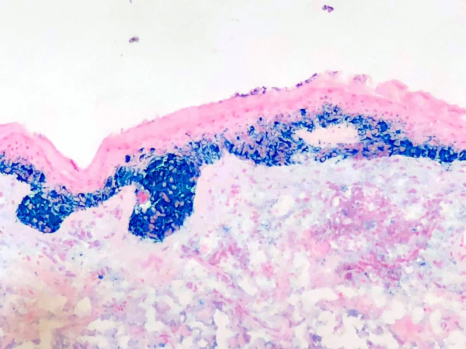
We perform same-day Mohs surgery for melanoma with immunohistochemical staining — MART-1 and SOX10 — run in our own laboratory during your surgery. These stains make melanoma cells visible at the margin in a way routine frozen sections cannot. The cancer is removed, every margin is tested with the stains, and once the margins are clear the wound is reconstructed — all on the same day. The traditional alternatives are wide local excision, or a staged excision (sometimes called "slow Mohs") that stretches the removal and margin reading over one to three days. True same-day, margin-controlled Mohs for melanoma is what this laboratory was built to do.
- MART-1 and SOX10 immunostains, run in our own laboratory
- Removal, margin testing, and reconstruction in a single day
- 100% of the margin examined for melanoma and melanoma in situ
- Tissue-sparing alternative to wide local excision
- No multi-day staged excision ("slow Mohs") — margins are read while you wait
What Sets Us Apart
Immunostaining for High-Risk Skin Cancers
Aggressive non-melanoma tumors traced accurately to their true margins.
The same on-site immunostaining capability is applied to aggressive non-melanoma tumors, including high-risk squamous cell carcinoma. Cancers that are difficult to see on routine sections can be traced accurately to their margins, which makes them amenable to tissue-sparing surgery with higher cure rates.
- Cytokeratin immunostains (AE1/AE3) for high-risk squamous cell carcinoma and extramammary Paget's disease
- Same-day, on-site processing
- Margin control for poorly-defined tumors
Skilled Reconstruction
Reconstructive Surgery
Expert wound closure and reconstruction after cancer removal.
After skin cancer removal, our surgeons provide expert reconstructive techniques to restore both function and appearance. We work to achieve optimal cosmetic results while ensuring complete cancer removal.
- Wound closure with stitches
- Skin flap reconstruction
- Skin graft procedures
- Healing by secondary intention when appropriate
- Minimized scarring techniques
- Coordination with other specialists when needed
Your Mohs surgeons
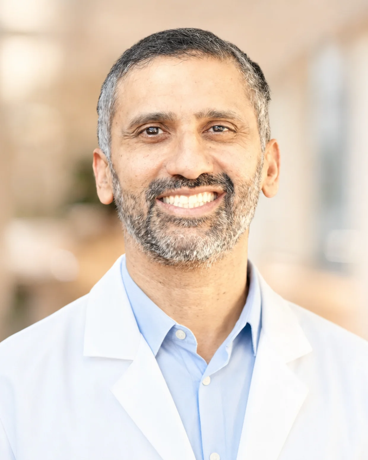
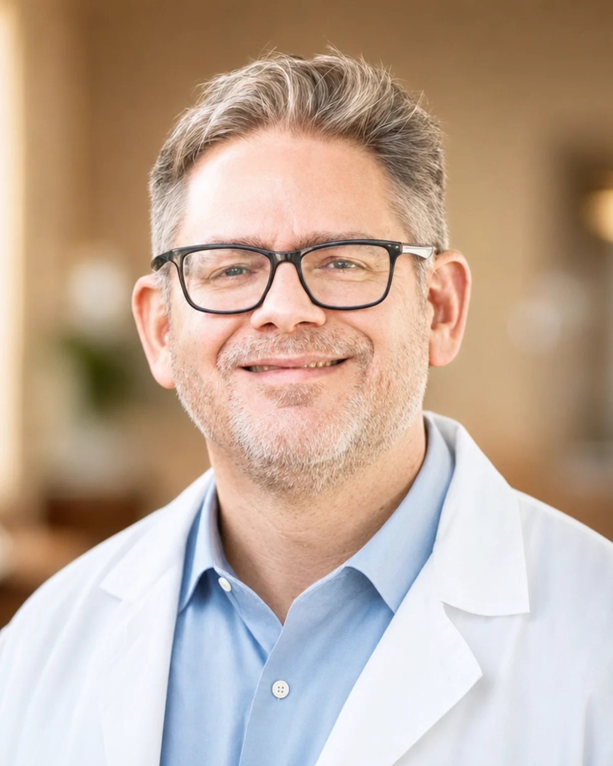
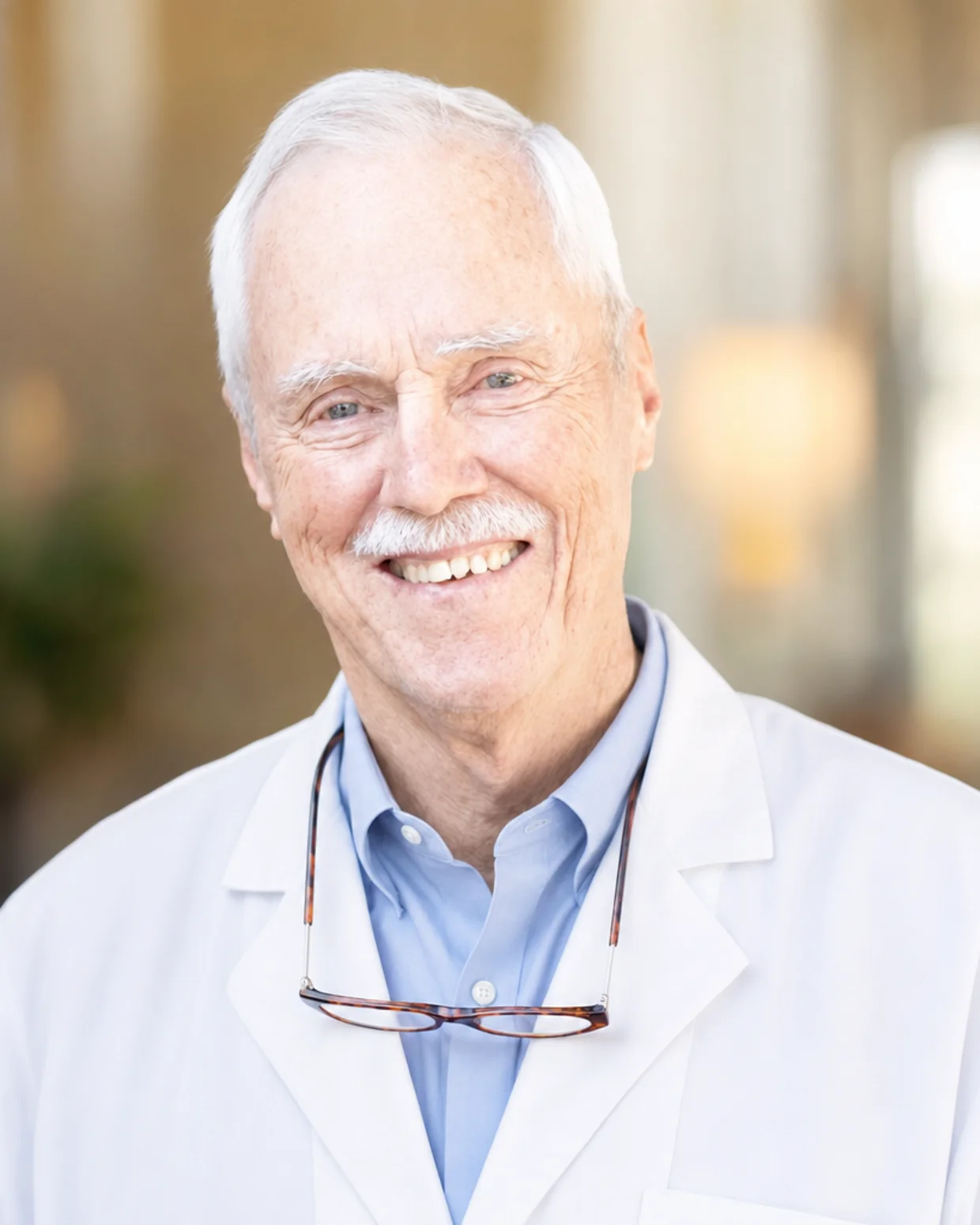
Each is board certified in dermatology and fellowship trained in Mohs surgery. Between them, they have trained fifteen Mohs surgeons and hundreds of dermatology residents.
Common questions about Mohs surgery
For anything else, call the office — you will reach a person, not a phone tree.
(972) 378-0620Planning your visit
Can Mohs Surgery Be Used for Melanoma?
Yes, for selected melanomas. Here is how Mohs compares with wide excision, what the recurrence and survival studies show, and who may be a candidate.
Read the articleTraveling to Plano
Patients travel to us from across North Texas — Frisco, Allen, McKinney, Richardson, Dallas, Sherman, Denison, Denton — and from southern Oklahoma. Our office is on Plano's west side, minutes from the Dallas North Tollway and the Sam Rayburn Tollway.
Areas we serveReferring physicians
Most referred patients are seen within days. We report back promptly after surgery, and we return every patient to your care.
How to refer a patientContact and directions
6100 Windhaven Parkway, Plano, TX 75093. Monday-Thursday: 7:30am - 4:00pm. Friday: 10:00am - 2:00pm.
Contact the officeLooking for something other than skin cancer surgery? See everything we treat.
Request a Consultation
Talk to us about your diagnosis
Most patients are seen within days of referral.
(972) 378-06206100 Windhaven Parkway, Plano, TX 75093 · Mon-Thurs: 7:30-4 | Fri: 10-2