Can Mohs Surgery Be Used for Melanoma?
Medically reviewed by Gunjan Modi, MD, FAAD, FACMS, board-certified dermatologist and fellowship-trained Mohs surgeon · Updated September 13, 2026
The short answer
Yes. Mohs surgery is an established option for melanoma in situ, especially the lentigo maligna type on the face and scalp, and experienced teams also use it for thin invasive melanomas. At this practice we offer same-day Mohs for appropriate cases of both. Mohs is designed to examine the complete outer and deep edges of the removed tissue, using special stains that make pigment cells visible. Published studies report low rates of melanoma returning at the treated spot, and some large registry studies have found better survival among patients treated with Mohs, though none of them can prove Mohs caused the difference.
How melanoma surgery usually works
The standard operation for melanoma is wide local excision. The surgeon removes the biopsy site and any remaining melanoma together with a measured border of normal-looking skin. For invasive melanoma that border is usually one to two centimeters, roughly three-eighths to three-quarters of an inch, depending on how thick the melanoma is. Melanoma in situ usually starts with a smaller planned margin, though some need more skin removed to clear their edges.
The tissue goes to a pathology laboratory, where sections are examined to describe the tumor and check whether it reaches the edges of the specimen. Results usually take several days. Wide excision is the guideline-recommended standard for invasive melanoma and works well for many patients. Standard pathology samples the edges rather than examining the complete outer and deep margins the way Mohs does. If melanoma is found at an edge, another operation may be needed.
How Mohs surgery for melanoma is different
During Mohs, the surgeon removes the tissue in carefully mapped stages. The outer and deep edges of each stage are prepared and examined under the microscope while you wait. If melanoma is found at an edge, the surgeon removes another thin layer from exactly that spot and checks again. Repair is planned once the examined margins are clear.
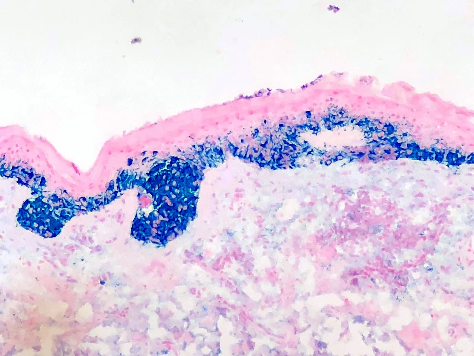
Melanoma Mohs needs specialized laboratory methods. Special stains, MART-1 and SOX10, highlight melanocytes, the pigment cells melanoma arises from. The stains highlight normal pigment cells too, so the surgeon reads the pattern, the number, and the arrangement of the stained cells to judge whether melanoma is present. Our laboratory runs these stains in-house during your surgery, which is what makes same-day margin assessment possible. Appropriate cases have removal and repair in the same visit; some need additional testing or a different repair plan.
Checking mapped margins also helps preserve healthy skin while the melanoma is removed. That matters most near the eyes, nose, lips, and ears, where extra tissue removal affects function and repair. The central tumor tissue is examined separately to confirm depth and the other features that guide staging. Occasionally this shows a higher stage than the original biopsy suggested and changes the plan. In one published Mohs series, 5.5 percent of melanomas were upstaged this way, and almost all were identified before repair.
Recurrence: what the numbers show
Many studies of melanoma Mohs focus on local recurrence, meaning melanoma returning at the treated spot. A 2022 systematic review pooled 71 published studies covering 16,575 patients. The pooled local recurrence estimates were about 7 percent after wide local excision, 3 percent after staged excision, and less than 1 percent after Mohs. Staged excision also uses mapped margins, usually with laboratory processing that takes longer. The studies included different tumors and follow-up periods, so these figures are a pattern across the literature, not a direct comparison of otherwise identical patients.
Several individual Mohs series report low local recurrence too, although they have no wide-excision group to compare against. In a multicenter study of 785 invasive melanomas of the head and neck treated with Mohs and MART-1 staining, four local recurrences were reported, about half of one percent, over a twelve-year study period. Another series reported seven local recurrences among 1,419 primary melanomas, again about half of one percent. In a separate single-center study of melanoma in situ, estimated five-year recurrence was 1.1 percent after Mohs and 4.1 percent after wide excision, a difference that was not statistically conclusive.
Figure
Local recurrence reported across published studies
Melanoma in situ and invasive melanoma combined; follow-up times varied.
Wide local excision
Staged excision
Mohs surgery
Pride and colleagues, Dermatologic Surgery, 2022, pooled 71 studies involving 16,575 patients. The studies differed in melanoma type, location, treatment methods, and length of follow-up. These are pooled local-recurrence estimates, not results from one randomized comparison and not rates measured at one common time point. Local recurrence means melanoma returning at the treated site; it does not measure all spread or deaths. Thin lines show 95% confidence intervals. The hatched band marks the reported ceiling for Mohs (“less than 1%”), not an exact value.
See the numbers as a table
| Technique | Local recurrence | 95% confidence interval |
|---|---|---|
| Wide local excision | 7% | 5–11% |
| Staged excision | 3% | 2–4% |
| Mohs surgery | less than 1% | 0–1% |
Survival: the newest evidence
Keeping melanoma from returning at the treated spot is important, but it is not the same as preventing spread or death. Survival comparisons between Mohs and wide excision come from observational studies, which can show an association but cannot prove that the operation caused a difference.
A 2026 study used the U.S. SEER cancer registry to compare melanoma-specific survival after Mohs and after wide excision in more than 90,000 patients with invasive melanoma treated between 2000 and 2020. The reported five-year melanoma-specific survival was 93 percent after Mohs and 88 percent after wide excision. At ten years it was 88 percent and 83 percent. These figures count deaths attributed to melanoma, not deaths from all causes, and they were not adjusted for the differences between the two groups. The authors also reported that an association favoring Mohs remained after statistical adjustment, but adjustment cannot make the groups fully comparable.
Figure
Reported melanoma-specific survival estimates
One retrospective registry study of more than 90,000 patients with invasive melanoma (U.S. SEER, 2000–2020). The treatment groups differed before surgery.
- Mohs surgery
- Wide local excision
Reported 5- and 10-year estimates from Sharma and colleagues, Archives of Dermatological Research, 2026. Dashed lines connect the reported points; they are not the study’s survival curves. These estimates were not adjusted for differences between the groups: patients selected for Mohs generally had thinner, earlier-stage melanomas. The results do not show that Mohs caused better survival. Melanoma-specific survival is not the same as survival from all causes.
See the numbers as a table
| Years after diagnosis | Mohs surgery | Wide local excision |
|---|---|---|
| At diagnosis | 100% | 100% |
| 5 years | 93% | 88% |
| 10 years | 88% | 83% |
The same estimates, shown with 100 squares
The display below shows the same survival estimates in another form. Each square is one percentage point, not an individual patient. The gap is five percentage points at both time points. That does not tell us how many lives either operation saved.
Figure
The same estimates, shown with 100 squares
Each square is one percentage point of the reported estimate, not one patient followed in the study.
At 5 years
Mohs surgery93%
Wide local excision88%
At 10 years
Mohs surgery88%
Wide local excision83%
Filled: estimated melanoma-specific survivalOutline: remaining share to 100%
These squares illustrate the same estimates as the chart above (Sharma et al., 2026); they are not additional study results. Deaths from other causes are not counted as melanoma deaths, so the percentages do not say how many people were still alive overall. The groups differed before treatment, and the five-point gap does not mean Mohs saved five additional lives per 100 patients.
See the numbers as a table
| Mohs surgery | Wide local excision | Gap | |
|---|---|---|---|
| At 5 years | 93% | 88% | 5 points |
| At 10 years | 88% | 83% | 5 points |
What this study can and cannot tell you
This was a registry study, not a randomized trial, and the two groups were not the same to begin with. Patients treated with Mohs generally had thinner, earlier-stage tumors, more often on the head and neck, and more often of the lentigo maligna melanoma subtype. Statistical adjustment tries to account for the differences that were measured, but important differences can remain, including overall health and the other care each patient received. The authors describe their finding as a potential advantage that deserves confirmation in prospective studies, and we agree.
Other registry studies give useful context, though several drew on overlapping years of the same national database and should not be counted as fully independent confirmations. A 2019 study of stage I invasive melanoma and a 2020 study of head and neck melanoma both reported better overall survival among patients treated with Mohs. Overall survival includes deaths from all causes, so general health can influence those results. A 2021 study of melanoma on the trunk and limbs found no statistically significant survival difference between the two operations.
Taken together, these studies support considering Mohs for appropriately selected melanomas. They do not prove that Mohs is better than wide excision for survival, and they do not prove the two are equivalent. The choice depends on the melanoma's features, its location, what staging it needs, and the expertise available.
Who may be a candidate
Mohs may be considered when a melanoma's borders are hard to define or when preserving nearby healthy skin matters. These are reasons for an assessment, not automatic eligibility:
- Melanoma in situ, including lentigo maligna, especially on the face, scalp, ears, and neck, where the cancer often extends beyond its visible edge
- Thin invasive melanomas in areas where preserving tissue helps protect function or simplify repair, after reviewing tumor depth and any need for lymph node evaluation
- Some melanomas with tumor remaining at a previous surgical margin, or a local recurrence, after the diagnosis and stage have been reassessed
- Melanomas with poorly defined borders
When wide excision is the right choice
Wide local excision remains the standard operation for invasive melanoma and is often the most appropriate approach, including for many melanomas on the trunk and limbs. Tumor thickness and other pathology findings determine whether a sentinel lymph node biopsy should be discussed. If melanoma has already been found in a lymph node, that calls for a separate staging and treatment plan. We recommend, or coordinate, whatever care your melanoma needs.
The decision weighs tumor thickness, ulceration and other pathology findings, location, lymph node evaluation, your health, and your preferences. Your surgeon will explain the recommendation and bring in another specialist when that is the right thing to do.
This page is general education, not medical advice for your specific situation. Published study results are not a prediction or a guarantee of your outcome; the right operation for a melanoma depends on its depth, its location, and your health. If you have a new diagnosis and want a straight answer about your options, call us at (972) 378-0620.
Common questions
Answers reflect the general case — a physician who can see the wound always beats a page that cannot.
Selected peer-reviewed literature
The data behind the answer.
- Sharma D, Taylor MA, Thomas S, Farberg A, Voss VB. Improved disease-specific outcomes in invasive cutaneous melanomas treated with Mohs surgery compared to wide local excision. Archives of Dermatological Research. 2026;318:242. (opens in new tab)
- Pride RLD, et al. Local recurrence of melanoma is higher after wide local excision versus Mohs micrographic surgery or staged excision: a systematic review and meta-analysis. Dermatologic Surgery. 2022;48(2):164–170. (opens in new tab)
- Cheraghlou S, et al. Comparison of survival after Mohs micrographic surgery vs wide margin excision for early-stage invasive melanoma. JAMA Dermatology. 2019;155(11):1252–1259. (opens in new tab)
- Hanson J, et al. Improved overall survival of melanoma of the head and neck treated with Mohs micrographic surgery versus wide local excision. Journal of the American Academy of Dermatology. 2020;82(1):149–155. (opens in new tab)
- Demer AM, et al. Association of Mohs micrographic surgery vs wide local excision with overall survival outcomes for patients with melanoma of the trunk and extremities. JAMA Dermatology. 2021;157(1):84–89. (opens in new tab)
- Beal BT, et al. Outcomes of invasive melanoma of the head and neck treated with Mohs micrographic surgery: a multicenter study. Journal of the American Academy of Dermatology. 2023;89(3):544–550. (opens in new tab)
- Valentín-Nogueras SM, et al. Mohs micrographic surgery using MART-1 immunostain in the treatment of invasive melanoma and melanoma in situ. Dermatologic Surgery. 2016;42(6):733–744. (opens in new tab)
- Etzkorn JR, et al. Low recurrence rates for in situ and invasive melanomas using Mohs micrographic surgery with MART-1 immunostaining: tissue processing methodology to optimize pathologic staging and margin assessment. Journal of the American Academy of Dermatology. 2015;72(5):840–850. (opens in new tab)
- Nosrati A, et al. Outcomes of melanoma in situ treated with Mohs micrographic surgery compared with wide local excision. JAMA Dermatology. 2017;153(5):436–441. (opens in new tab)
- Stigall LE, Brodland DG, Zitelli JA. The use of Mohs micrographic surgery for melanoma in situ of the trunk and proximal extremities. Journal of the American Academy of Dermatology. 2016;75(5):1015–1021. (opens in new tab)
- Swetter SM, et al. Guidelines of care for the management of primary cutaneous melanoma. Journal of the American Academy of Dermatology. 2019;80(1):208–250. (opens in new tab)
If you have been diagnosed with melanoma
Removing skin cancer with the margins checked is what this practice does all day, every day. We treat melanoma and the other common skin cancers, and we will tell you plainly which operation fits yours.
Mohs Surgery for Melanoma
The most serious common skin cancer — treated here with same-day, margin-controlled Mohs surgery for appropriate cases.
Basal Cell Carcinoma
The most common skin cancer — slow to spread, but destructive locally. Mohs offers the most precise removal.
Squamous Cell Carcinoma
The second most common skin cancer — treated promptly because it can spread. High-risk tumors get immunostained margin control.
Request a Consultation
A question about a spot that worries you?
Call us, or send an appointment request — requests receive a call back within one business day.
(972) 378-06206100 Windhaven Parkway, Plano, TX 75093 · Mon-Thurs: 7:30-4 | Fri: 10-2