My Biopsy Says ‘Atypical Mole.’ Is That Cancer?
Medically reviewed by Gunjan Modi, MD, FAAD, FACMS, board-certified dermatologist and fellowship-trained Mohs surgeon · Updated October 3, 2026
The short answer
No. An atypical mole, also called a dysplastic nevus, is not melanoma. It is a mole whose cells look unusual under the microscope. Most never become melanoma, and about 7 in 10 melanomas show no sign of a mole under the microscope. There is no percentage for how many atypical moles turn into melanoma; that number does not exist in the research. Your next step depends on two things in your report: the grade, mild, moderate, or severe, and whether mole cells reach the edge of the sample. Mild and moderate moles with clear edges usually need no more surgery. Removing more skin is discussed more often for severe atypia or cells at the edge. Atypical moles can also signal a higher melanoma risk elsewhere, so keep up skin checks. Your dermatologist makes the plan with you.
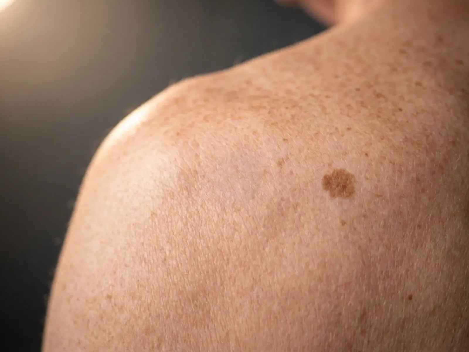
What “atypical mole” and “dysplastic nevus” mean
Atypical mole and dysplastic nevus are two names for the same finding. A nevus is a mole. Dysplastic, or atypical, means that under the microscope the mole's cells and their pattern look more unusual than those of an ordinary mole. Your report may use other names for it: atypical nevus, nevus with architectural disorder and cytologic atypia or, under the newer World Health Organization system, low-grade or high-grade dysplastic nevus. They all describe the same kind of mole.
It is not melanoma. The pathologist who read your slide was looking for melanoma and did not find it; that is why the report says nevus. The National Cancer Institute's patient page puts it this way: only rarely does a dysplastic nevus turn into melanoma. The report matters, as the rest of this page explains, but it is not a cancer diagnosis.
Two things are easy to mix up. A mole can be called atypical because of how it looks on your skin, and a mole can be called dysplastic because of how it looks under the microscope. This page is about the second one: a biopsy report whose diagnosis is a dysplastic or atypical nevus. If your report uses words like “cannot exclude melanoma” or “atypical melanocytic proliferation,” it is describing an uncertain finding, and that is a different conversation to have with your dermatologist.
Mild, moderate, severe: what the grade means, and why wording differs
Most reports grade the atypia as mild, moderate, or severe. Some labs use two grades instead, low-grade and high-grade. The grade is the pathologist's judgment of how far the cells and their pattern stray from an ordinary mole. It is not a cancer stage. There is no blood test or measurement behind it. It is a trained eye looking at a stained slide.
Because it is a judgment, it varies. In the largest study of this question, 187 pathologists from ten states each read a set of slides drawn from 240 melanocytic biopsy cases. Counting all the readers together, an average of ten different diagnostic terms were applied to each case. Agreement with a panel of experienced pathologists was high for ordinary moles and mild atypia and for thicker melanomas, and lowest for moderate atypia. The chart below shows the numbers.
Severe atypia deserves its own sentence. It is still not melanoma. But in that study, severe atypia and melanoma in situ, the earliest form of melanoma, were grouped in the same class, because on a slide the two can be hard to tell apart. A study of severely atypical moles says the same thing: they are often re-excised out of concern that the lesion may in fact be an early melanoma. That resemblance, not a proven path from one to the other, is why severe atypia is handled more cautiously, as the sections below explain.
Figure
How often a pathologist's reading matched an expert panel
A study in which 187 U.S. pathologists each read a set of 36 or 48 slides drawn from 240 biopsy cases. Each bar is the share of their readings, by diagnostic class, that agreed with a consensus panel of experienced pathologists; the thin dark line is the 95% confidence interval.
Data: Elmore JG, et al. Pathologists’ diagnosis of invasive melanoma and melanocytic proliferations: observer accuracy and reproducibility study. BMJ. 2017;357:j2813. Phase 1 of the study: 8,976 independent readings. Each class groups several diagnoses; the names shown are the study’s own examples. The panel is a reference standard, not a perfect answer. For a typical mix of cases in everyday practice, the same authors estimated that 82.8% (81.0% to 84.5%) of melanocytic biopsy diagnoses would be confirmed by such a panel. Class III groups severe atypia together with melanoma in situ, so that bar is not severe atypical moles alone.
What this means: the middle grades are where wording differs most from one pathologist to another. That is why a dermatologist sometimes asks a second pathologist to read a slide. This chart is about agreement between pathologists. It does not mean your report is wrong, and it is not the chance that your mole is cancer. This figure is our own drawing of the published numbers.
See the numbers as a table
| Diagnostic class | Agreed with the panel | 95% confidence interval |
|---|---|---|
| Ordinary mole or mild atypia (class I) | 92% | 90% to 94% |
| Moderate atypia (class II) | 25% | 22% to 28% |
| Severe atypia or melanoma in situ (class III) | 40% | 37% to 44% |
| Early invasive melanoma (class IV, stage pT1a) | 43% | 39% to 46% |
| Invasive melanoma, stage pT1b or higher (class V) | 72% | 69% to 75% |
Does an atypical mole turn into melanoma?
Here is the honest answer: no study gives a percentage for how often a dysplastic nevus becomes melanoma, and any such number you read online was not measured. To measure it, researchers would have to leave atypical moles in place and watch them for decades. Once a mole has been biopsied, the question changes. What the research does give is a set of indirect answers, and they all point the same way.
First, any single mole, atypical or not, has a very small chance of becoming melanoma. A 2003 model of all moles in the U.S. population put the yearly chance for one mole at 0.0005 percent or less (1 in 200,000 or fewer) for people under 40, rising to about 0.003 percent a year (about 1 in 33,000) for men over 60. That is a model of moles in general, not a count of dysplastic ones, and it is older data. Second, most melanomas show no sign of a mole. A 2017 analysis of 38 studies found that 70.9 percent of melanomas, about 7 in 10, had no trace of a mole under the microscope, meaning they most likely arose on their own. Among the melanomas that did arise in a mole, it found no significant difference between dysplastic and ordinary moles as the starting point.
Third, when atypical moles that were biopsied but not fully removed have been followed, melanoma at the biopsy site has not turned up. In a study from nine academic centers, 467 moderately dysplastic nevi with positive margins were watched for an average of 6.9 years, and none developed into melanoma at the site. At one center, 115 dysplastic nevi that reached or nearly reached the edge of the biopsy were followed for an average of 17.4 years, though not every patient was followed that long, with the same result. Zero in a study is not the same as zero risk, these were patients under regular skin checks, and almost all of the moles were mild or moderate, so these studies say little about severe atypia. Whether these moles are a step toward melanoma or only a marker of risk is still argued among experts. What the best data say is that the risk at the site of a biopsied mild or moderate atypical mole is very low.
Why the report still matters: a marker of your overall risk
The reason dermatologists care about atypical moles is not that one mole is likely to turn. It is that people who have many of them are more likely to develop melanoma somewhere on their skin. A meta-analysis of 46 studies found that people with 5 atypical moles had 6.36 times the melanoma risk of people with none (95 percent confidence interval 3.80 to 10.33). Having many ordinary moles counted too: 101 to 120 moles versus fewer than 15 carried 6.89 times the risk (4.63 to 10.25). A companion analysis of 60 studies found that a family history of melanoma carried 1.74 times the risk (1.41 to 2.14).
Three cautions. These counts come from moles examined on the skin, not from biopsy reports, so one biopsy that reads “atypical” is not the same as having five atypical moles. The numbers are relative, meaning how risk compares between groups, not your personal odds. And they come from studies published before 2002. They still carry the practical message: the more atypical moles you have, and the more melanoma in your family, the more your whole skin needs watching.
The 467-mole study above makes the same point from the other side. Its patients came from academic clinics that see many high-risk people, and 22.8 percent of them were later found to have a melanoma somewhere else on the skin. By far the strongest predictor was having already had a melanoma (odds ratio 11.74). If that is you, our article I Just Had a Melanoma. What Are My Odds of Getting Another One? covers what that history means for your follow-up.
What a positive margin means, and what the consensus says
A biopsy is taken to make a diagnosis, not always to remove the whole mole, so it is common for the report to say the atypical cells reach the edge of the sample. That is a “positive margin.” In one series of 1,809 mild and moderate dysplastic nevi, 42.3 percent had a positive margin; in another series of 580 dysplastic nevi, 34 percent did, more often as the grade went up. A positive margin is a normal result of how shave and punch biopsies work. It does not mean anything went wrong, and it does not mean the mole has spread.
What happens next depends on the grade and the margin. In 2015 a national panel of pigmented-lesion specialists reached consensus on three points: mild and moderate dysplastic nevi with clear margins do not need to be removed again; a mild one with a positive margin and no visible pigment left at the site can be safely watched; and watching may be reasonable for a moderate one with a positive margin and no visible pigment left, though the panel said more data were needed before a firm recommendation. The panel reached its view by structured expert agreement, not a trial, and it did not address severe atypia. The 467-mole study and the 115-mole study above are the kind of data the panel asked for, and both support watching.
That is what many dermatologists do. It is not a rule for your mole. Notice the condition in the consensus: no visible pigment left at the site. A positive margin under the microscope is different from mole color you can still see in or around the scar, and the watching evidence covers only moles with none, so tell your dermatologist about any leftover color. If pigment is still visible, if the reading was borderline, or if your history raises concern, your dermatologist may recommend removing more. Surgeons differ here, and the evidence allows it.
What re-excision usually finds, and the severe grade
Removing more skin around a mild or moderate atypical mole rarely changes anything. In one series, 495 such moles with positive margins were re-excised: leftover mole cells were found in 18.2 percent, and the diagnosis changed in a clinically meaningful way in 1 case (0.2 percent). That study measured what re-excision finds; its follow-up was too short to say anything about how a mole behaves over the years. When moderately atypical moles with positive margins were left alone in another series, 6 of 147 (4 percent) grew back, all after shave biopsies, on average 1.7 years later. Regrowth is the mole coming back, not melanoma, but it is one reason the scar is worth watching.
Severe atypia is handled differently, and the research explains why. In one laboratory's series of 580 dysplastic nevi, 127 with positive margins were re-excised, and in 2 of them (1.6 percent) the diagnosis changed to melanoma in situ; both had been read as moderately to severely atypical on the biopsy. In a single-center study of 451 patients with severely dysplastic nevi, 36.6 percent had a re-excision, and 2 melanomas were found in those specimens. Those authors concluded that re-excising every severe lesion may not be necessary, while the authors of a different study wrote that clinicians agree a severe lesion with a positive margin should be removed with a margin. In other words, many dermatologists remove a little more skin around a severely atypical mole, some watch closely when the margins are clear, and both are defensible.
A re-excision of an atypical mole is a standard excision with stitches, not Mohs surgery. If a re-excision does find melanoma in situ, treatment follows the melanoma path; see our melanoma page and Can Mohs Surgery Be Used for Melanoma?. Whatever your report says, your own dermatologist will weigh the grade, the margin, your skin exam, and your history with you.
Watching your skin from here
An atypical mole on a report is a reason to keep up with skin checks, not a reason to worry every day. The National Cancer Institute's patient page suggests checking your own skin monthly, a yearly skin exam by a doctor for people with more than five dysplastic nevi, and more frequent exams when there is a family history of melanoma. Those are general suggestions. Your dermatologist sets your schedule based on your mole count, your family history, and whether you have had melanoma before.
Two habits help. Watch the biopsy scar: if pigment comes back in or around it, show your dermatologist, because a regrown mole can look worrying under the microscope and it helps the pathologist to know there was a biopsy there. And tell any new doctor about this biopsy so it is in your record. Protect your skin from the sun with shade, clothing, and a broad-spectrum sunscreen; sun exposure is the one part of melanoma risk you control.
If your biopsy site is still healing, follow the wound-care instructions from the clinician who did the biopsy.
Questions to bring to your appointment
A pathology report is easier to live with once you know what it means for you. These questions cover the points this page cannot answer for your mole:
- What grade did the pathologist give, and did the cells reach the edge of the sample?
- Is there any pigment left at the biopsy site?
- Do you recommend watching it or removing more, and why?
- Would a second pathologist's reading of the slide change the plan?
- How many atypical moles do I have, and does my family history change my risk?
- How often should I have a full skin exam, and should I take photos of my moles?
This page is general education, not medical advice about your biopsy report. Published study results are averages across many people and are not a prediction for your mole; what happens next depends on the grade, the margins, your skin exam, and your history. If you have a report in hand and want a plain explanation of your options, call us at (972) 378-0620.
Common questions
Answers reflect the general case — a physician who can see the wound always beats a page that cannot.
Selected peer-reviewed literature
The data behind the answer.
- Elmore JG, et al. Pathologists' diagnosis of invasive melanoma and melanocytic proliferations: observer accuracy and reproducibility study. BMJ. 2017;357:j2813. (opens in new tab)
- Shea CR, Prieto VG, Shachaf CM, Florell SR. Grading melanocytic dysplasia: updated histopathologic criteria. Journal of Cutaneous Pathology. 2025;53(1):29–37. (opens in new tab)
- Tsao H, Bevona C, Goggins W, Quinn T. The transformation rate of moles (melanocytic nevi) into cutaneous melanoma: a population-based estimate. Archives of Dermatology. 2003;139(3):282–288. (opens in new tab)
- Pampena R, et al. A meta-analysis of nevus-associated melanoma: prevalence and practical implications. Journal of the American Academy of Dermatology. 2017;77(5):938–945. (opens in new tab)
- Gandini S, et al. Meta-analysis of risk factors for cutaneous melanoma: I. Common and atypical naevi. European Journal of Cancer. 2005;41(1):28–44. (opens in new tab)
- Gandini S, et al. Meta-analysis of risk factors for cutaneous melanoma: III. Family history, actinic damage and phenotypic factors. European Journal of Cancer. 2005;41(14):2040–2059. (opens in new tab)
- Kim CC, et al. Addressing the knowledge gap in clinical recommendations for management and complete excision of clinically atypical nevi/dysplastic nevi: Pigmented Lesion Subcommittee consensus statement. JAMA Dermatology. 2015;151(2):212–218. (opens in new tab)
- Kim CC, et al. Risk of subsequent cutaneous melanoma in moderately dysplastic nevi excisionally biopsied but with positive histologic margins. JAMA Dermatology. 2018;154(12):1401–1408. (opens in new tab)
- Hocker TL, Alikhan A, Comfere NI, Peters MS. Favorable long-term outcomes in patients with histologically dysplastic nevi that approach a specimen border. Journal of the American Academy of Dermatology. 2013;68(4):545–551. (opens in new tab)
- Strazzula L, et al. The utility of re-excising mildly and moderately dysplastic nevi: a retrospective analysis. Journal of the American Academy of Dermatology. 2014;71(6):1071–1076. (opens in new tab)
- Reddy KK, Farber MJ, Bhawan J, Geronemus RG, Rogers GS. Atypical (dysplastic) nevi: outcomes of surgical excision and association with melanoma. JAMA Dermatology. 2013;149(8):928–934. (opens in new tab)
- Hiscox B, et al. Recurrence of moderately dysplastic nevi with positive histologic margins. Journal of the American Academy of Dermatology. 2017;76(3):527–530. (opens in new tab)
- Engeln K, et al. Dysplastic nevi with severe atypia: long-term outcomes in patients with and without re-excision. Journal of the American Academy of Dermatology. 2017;76(2):244–249. (opens in new tab)
- National Cancer Institute. Common Moles, Dysplastic Nevi, and Risk of Melanoma (patient fact sheet). (opens in new tab)
If you need a mole removed, or your report raises the question of melanoma
Removing moles and skin cancers with the margins checked is the daily work of this practice. If your dermatologist recommends a re-excision, or a report raises the question of melanoma, we will tell you plainly what the next step is.
Mohs Surgery for Melanoma
The most serious common skin cancer — treated here with same-day, margin-controlled Mohs surgery for appropriate cases.
Mole Evaluation & Removal
Surgical evaluation and removal of irregular, symptomatic, or unwanted moles — with pathology when it matters.
Request a Consultation
A question about a spot that worries you?
Call us, or send an appointment request — requests receive a call back within one business day.
(972) 378-06206100 Windhaven Parkway, Plano, TX 75093 · Mon-Thurs: 7:30-4 | Fri: 10-2